How does the endoscopy camera system’s calibration work?
As a supplier of Endoscopy Camera Systems, I’ve witnessed firsthand the crucial role calibration plays in the performance…
Read article →

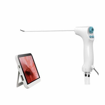
Endometrial hyperplasia is a condition characterized by the abnormal thickening of the endometrium, the inner lining of the uterus. It is often associated with an imbalance in the hormones estrogen and progesterone, and if left untreated, it can potentially progress to endometrial cancer. Accurate and timely diagnosis of endometrial hyperplasia is crucial for appropriate management and treatment. One of the most effective diagnostic tools for this condition is the hysteroscope. As a leading hysteroscope supplier, we understand the significance of this technology in the field of gynecology. In this blog, we will explore how a hysteroscope helps in diagnosing endometrial hyperplasia.


Hysteroscopy is a minimally invasive procedure that allows direct visualization of the inside of the uterus. A hysteroscope is a thin, lighted tube equipped with a camera that is inserted through the vagina and cervix into the uterus. The camera transmits real – time images of the uterine cavity to a monitor, enabling the gynecologist to examine the endometrium closely. There are different types of hysteroscopes available in the market, each with its own unique features and benefits. For instance, the Flexible Hysteroscope offers greater flexibility, allowing it to navigate through the curved anatomy of the uterus more easily. The Portable Hysteroscope provides convenience as it can be used in various settings, including outpatient clinics. And the Single Use Hysteroscope eliminates the risk of cross – contamination.
The primary way a hysteroscope aids in diagnosing endometrial hyperplasia is by providing direct visualization of the endometrium. In normal circumstances, the endometrium appears as a smooth, thin layer lining the uterine cavity. However, in cases of endometrial hyperplasia, the hysteroscope can reveal characteristic changes. The hyperplastic endometrium may appear thickened, with an irregular or lumpy texture. There may be areas of polyps or abnormal growths within the uterine cavity. These visual cues are essential for the initial assessment of the condition. Unlike other diagnostic methods such as ultrasound, which provides a more indirect view of the uterus, hysteroscopy offers a real – time, high – resolution image of the endometrium. This allows the gynecologist to accurately identify any abnormal areas and determine the extent of the hyperplasia.
In addition to visual inspection, hysteroscopy also enables targeted biopsy. A biopsy is the removal of a small sample of tissue from the endometrium for laboratory examination. This is a critical step in confirming the diagnosis of endometrial hyperplasia and determining its type (e.g., simple hyperplasia, complex hyperplasia, with or without atypia). With the help of the hysteroscope, the gynecologist can precisely locate the abnormal areas within the uterine cavity and obtain tissue samples directly from those sites. This targeted approach ensures that the biopsy specimen is representative of the病变, leading to more accurate diagnosis and prognosis. For example, in cases where the hyperplasia is focal, meaning it is only present in specific areas of the endometrium, a targeted biopsy using hysteroscopy is far more effective than a random sampling method.
Endometrial hyperplasia can sometimes present with symptoms similar to other uterine conditions, such as uterine fibroids, polyps, or endometrial cancer. Hysteroscopy plays a vital role in differentiating between these conditions. By visualizing the uterine cavity, the gynecologist can observe the characteristics of the lesions. For example, uterine fibroids typically appear as well – defined, solid masses, while endometrial hyperplasia shows as a thickened and often irregular endometrial layer. Furthermore, the ability to perform a biopsy during hysteroscopy allows for histological examination, which can definitively distinguish between hyperplasia and cancer. The presence of abnormal cells under the microscope, such as abnormal cell growth patterns or nuclear atypia, can help in making a precise diagnosis.
Hysteroscopy also helps in assessing the severity of endometrial hyperplasia. The extent of the thickening of the endometrium, the presence of any associated polyps or tumors, and the degree of abnormal cell growth can all be evaluated during the procedure. Mild cases of endometrial hyperplasia may show only slight thickening of the endometrium, while more severe cases may present with extensive thickening, large polyps, or areas of atypical cells. This information is crucial for determining the appropriate treatment strategy. For mild hyperplasia, conservative treatment options such as hormone therapy may be sufficient, while more severe cases may require surgical intervention, such as hysterectomy.
The use of hysteroscopy in diagnosing endometrial hyperplasia ultimately leads to improved patient outcomes. Early and accurate diagnosis allows for timely treatment, which can prevent the progression of the condition to endometrial cancer. By providing a clear view of the uterine cavity and enabling targeted biopsy, hysteroscopy reduces the need for more invasive procedures in many cases. Patients can be spared from unnecessary surgeries or prolonged periods of undiagnosed illness. Moreover, the minimally invasive nature of hysteroscopy means less pain, shorter hospital stays, and faster recovery times for patients.
As a leading hysteroscope supplier, we are committed to providing high – quality hysteroscopes that meet the needs of gynecologists and patients alike. Our hysteroscopes are equipped with the latest technology, offering excellent image quality and easy maneuverability. The Flexible Hysteroscope is designed to provide a comfortable and efficient examination experience, even in complex anatomical situations. The Portable Hysteroscope is ideal for clinics and healthcare facilities that require flexibility and mobility. And the Single Use Hysteroscope ensures the highest level of patient safety.
If you are interested in learning more about our hysteroscopes or would like to discuss a potential purchase, we encourage you to reach out to us. We have a team of experts who are ready to provide you with detailed information and assist you in making the right choice for your practice.
 Blog
Blog
 Blog
Blog
 Blog
Blog