Top 10 Hysteroscope Manufacturers in the world
Introduction to Hysteroscope A hysteroscope is a thin, lighted tube that is inserted through the vagina and cervix…
Read article →

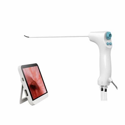
Müllerian duct anomalies (MDAs) are a group of congenital disorders that occur due to abnormal development of the Müllerian ducts during embryogenesis. These anomalies can lead to various gynecological and reproductive problems, such as infertility, recurrent pregnancy loss, and menstrual irregularities. Accurate diagnosis of MDAs is crucial for appropriate management and treatment. In recent years, flexible hysteroscopy has emerged as a potential diagnostic tool for MDAs. As a supplier of Flexible Hysteroscope, I will explore the feasibility and effectiveness of using a flexible hysteroscope to diagnose Müllerian duct anomalies in this blog.
The Müllerian ducts, also known as paramesonephric ducts, are paired embryonic structures that develop into the female reproductive tract, including the fallopian tubes, uterus, cervix, and upper part of the vagina. MDAs can result from complete or partial failure of the Müllerian ducts to develop, fuse, or canalize properly. According to the American Society for Reproductive Medicine (ASRM) classification system, MDAs are divided into seven main categories: agenesis or hypoplasia, unicornuate uterus, didelphys uterus, bicornuate uterus, septate uterus, arcuate uterus, and DES-related anomalies.


Each type of MDA has distinct anatomical features and clinical implications. For example, a septate uterus is characterized by a fibrous or muscular septum that divides the uterine cavity, which can increase the risk of recurrent pregnancy loss and preterm birth. On the other hand, a unicornuate uterus is a single-horned uterus that may be associated with a non-communicating rudimentary horn, which can cause pain and endometriosis.
Historically, several diagnostic methods have been used to evaluate MDAs, including pelvic ultrasound, hysterosalpingography (HSG), magnetic resonance imaging (MRI), and laparoscopy.
Pelvic ultrasound is a non-invasive and widely available imaging modality that can provide information about the size, shape, and structure of the uterus and ovaries. However, it has limitations in visualizing the internal uterine cavity and distinguishing between different types of MDAs.
HSG involves the injection of a contrast medium into the uterine cavity and fallopian tubes, followed by X-ray imaging. It can show the patency of the fallopian tubes and the outline of the uterine cavity. However, it is a invasive procedure and may not provide detailed information about the uterine septum or other subtle anatomical abnormalities.
MRI is a highly accurate imaging modality that can provide detailed anatomical information about the female reproductive tract. It can differentiate between different types of MDAs and is particularly useful for evaluating complex cases. However, it is expensive, time-consuming, and not readily available in all settings.
Laparoscopy is a surgical procedure that involves the insertion of a thin, lighted tube through a small incision in the abdomen to visualize the pelvic organs directly. It can provide a definitive diagnosis of MDAs and is often used in combination with hysteroscopy for treatment. However, it is an invasive procedure with potential risks and complications.
Flexible hysteroscopy is a minimally invasive procedure that involves the insertion of a thin, flexible scope through the vagina and cervix into the uterine cavity. The scope is equipped with a light source and a camera that allows direct visualization of the uterine cavity and the endometrial lining. Flexible Hysteroscope has several advantages over traditional diagnostic methods for MDAs.
One of the main advantages of flexible hysteroscopy is its ability to provide direct visualization of the uterine cavity. This allows for the accurate identification of anatomical abnormalities, such as uterine septa, polyps, fibroids, and adhesions. In the case of MDAs, flexible hysteroscopy can clearly show the presence and extent of a uterine septum, which is important for determining the appropriate treatment approach.
Flexible hysteroscopy allows for real-time assessment of the uterine cavity during the procedure. The operator can manipulate the scope to obtain different views and angles, which can help in differentiating between different types of MDAs. For example, a septate uterus can be distinguished from a bicornuate uterus by observing the shape and location of the uterine septum.
Compared to laparoscopy, flexible hysteroscopy is a minimally invasive procedure that can be performed in an outpatient setting without the need for general anesthesia. It has a shorter recovery time and fewer complications, making it a more patient-friendly option.
In addition to diagnosis, flexible hysteroscopy can also be used for therapeutic purposes. For example, it can be used to remove uterine polyps, fibroids, and adhesions, as well as to perform septum resection in cases of septate uterus. This combination of diagnostic and therapeutic capabilities makes flexible hysteroscopy a valuable tool in the management of MDAs.
Despite its many advantages, flexible hysteroscopy also has some limitations in diagnosing MDAs.
Flexible hysteroscopy is mainly focused on the uterine cavity and does not provide direct visualization of the external uterine surface or the pelvic organs. Therefore, it may not be able to detect certain types of MDAs, such as unicornuate uterus with a non-communicating rudimentary horn, which require evaluation of the extrauterine structures.
The accuracy of flexible hysteroscopy depends on the skill and experience of the operator. A less experienced operator may have difficulty in identifying subtle anatomical abnormalities or differentiating between different types of MDAs. Therefore, proper training and certification are essential for ensuring the quality of flexible hysteroscopy procedures.
Flexible hysteroscopy can be technically challenging, especially in cases of narrow cervical canals or uterine anomalies that distort the normal anatomy. In some cases, it may be necessary to use additional techniques, such as cervical dilation or the use of a guidewire, to facilitate the insertion of the scope.
To overcome the limitations of flexible hysteroscopy, it is often used in combination with other diagnostic methods. For example, pelvic ultrasound can be used as a preliminary screening tool to identify the presence of uterine abnormalities, followed by flexible hysteroscopy for further evaluation. MRI can also be used in complex cases to provide additional information about the extrauterine structures.
In addition, Disposable Hysteroscope can be used in combination with laparoscopy for a comprehensive evaluation of MDAs. Laparoscopy can provide direct visualization of the external uterine surface and the pelvic organs, while hysteroscopy can provide detailed information about the uterine cavity. This combined approach can improve the accuracy of diagnosis and guide the appropriate treatment.
Flexible hysteroscopy is a valuable diagnostic tool for Müllerian duct anomalies. It offers direct visualization of the uterine cavity, real-time assessment, and therapeutic potential, making it a minimally invasive and patient-friendly option. However, it also has some limitations, such as limited extrauterine visualization and operator dependence. To overcome these limitations, flexible hysteroscopy is often used in combination with other diagnostic methods.
As a supplier of flexible hysteroscopes, we are committed to providing high-quality products that meet the needs of healthcare professionals. Our flexible hysteroscopes are designed with advanced technology and features to ensure accurate diagnosis and effective treatment. If you are interested in learning more about our products or would like to discuss potential procurement opportunities, please feel free to contact us. We look forward to working with you to improve the diagnosis and management of Müllerian duct anomalies.
 Blog
Blog
 Blog
Blog