What is the signal transmission method of an endoscope camera system?
Hey there! As a supplier of Endoscope Camera Systems, I often get asked about how these nifty devices…
Read article →

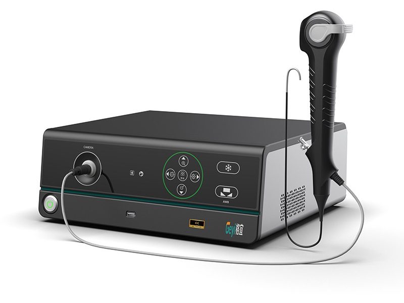
As a leading provider of 4K Laparoscopy Systems, we understand the critical role that image enhancement algorithms play in modern medical imaging. These algorithms are not just technical add – ons; they are the backbone of high – quality visualization during laparoscopic procedures, enabling surgeons to make more accurate diagnoses and perform complex operations with greater precision. In this blog, we will explore the various image enhancement algorithms used in our 4K Laparoscopy System.


One of the most fundamental aspects of image enhancement is contrast adjustment. In laparoscopic images, proper contrast is essential to clearly distinguish between different tissues, organs, and blood vessels.
Histogram equalization is a well – known contrast enhancement algorithm. It redistributes the intensity values of an image so that the histogram becomes more evenly distributed across the available range. This technique effectively stretches the contrast of the image, making details more visible. For example, in a laparoscopic view of the abdominal cavity, it can help to better differentiate between the pale – colored fat tissue and the darker – colored internal organs.
Adaptive histogram equalization (AHE) takes this concept further. Unlike traditional histogram equalization, which applies the same transformation to the entire image, AHE divides the image into small regions and performs histogram equalization on each region separately. This allows for local contrast enhancement, which is particularly useful in laparoscopic images where different parts of the field – of – view may have different lighting conditions. For instance, in a 4K Laparoscopy System, AHE can enhance the details in the shadowed areas near the edges of the surgical site while not over – enhancing the well – lit central areas.
Noise is an inevitable problem in medical imaging, including laparoscopic images. It can be caused by factors such as electronic interference in the camera sensor, low – light conditions, and the limitations of the image acquisition process. Noise can obscure important details and make it difficult for surgeons to accurately assess the surgical site.
Gaussian filtering is a commonly used noise reduction algorithm. It works by convolving the image with a Gaussian kernel, which is a matrix of weights that are based on the Gaussian distribution. The Gaussian filter smooths the image by averaging the pixel values in the neighborhood of each pixel. This reduces high – frequency noise while preserving the overall structure of the image. In our 4K Laparoscopy System, Gaussian filtering can be applied to the real – time video stream to provide a cleaner and more stable image for the surgeon.
Another powerful noise reduction technique is the non – local means algorithm. This algorithm takes advantage of the fact that similar patches of pixels can be found throughout the image. Instead of just using the local neighborhood of a pixel to estimate its value, the non – local means algorithm searches for similar patches in other parts of the image and uses their average value to replace the noisy pixel. This method can effectively reduce noise while maintaining fine details, which is crucial in laparoscopic surgery where even the smallest anatomical features can be significant.
Edges in an image represent the boundaries between different objects or tissues. Enhancing these edges can improve the visibility of anatomical structures and help surgeons to better understand the spatial relationships between different parts of the surgical site.
The Sobel operator is a simple yet effective edge detection and enhancement algorithm. It calculates the gradient of the image intensity in the horizontal and vertical directions. By combining these gradients, it can highlight the edges in the image. In a 4K Laparoscopy System, the Sobel operator can be used to enhance the boundaries of organs, blood vessels, and tumors, making them more distinct in the image.
The Canny edge detector is a more sophisticated edge enhancement algorithm. It consists of several steps, including Gaussian smoothing to reduce noise, gradient calculation, non – maximum suppression to thin the edges, and hysteresis thresholding to determine which edges are real and which are due to noise. The Canny edge detector can produce very accurate and clean edges, which are useful for tasks such as segmentation and measurement in laparoscopic images.
Color plays a vital role in laparoscopic imaging as it provides important information about the health and condition of tissues. For example, the color of the mucosa can indicate the presence of inflammation or disease.
White balance adjustment is a basic color enhancement technique. In a laparoscopic environment, the lighting conditions can vary significantly, which can cause color shifts in the image. White balance adjustment corrects these color shifts by ensuring that white objects in the image appear truly white. Our 4K Laparoscopy System is equipped with advanced white balance algorithms that can adapt to different lighting conditions in real – time, providing a more natural and accurate color representation.
Color constancy algorithms are also used to maintain consistent color perception across different lighting conditions. These algorithms attempt to estimate the color of the illumination source and then adjust the image colors accordingly. This is important in laparoscopic surgery where the color of tissues can be used to make important diagnostic decisions.
Image sharpening is used to enhance the clarity and definition of an image. It can make details more visible and improve the overall visual quality of the laparoscopic image.
Unsharp masking is a popular image sharpening algorithm. It works by creating a blurred version of the image and then subtracting it from the original image. The resulting difference image is then added back to the original image with a certain gain factor. This process enhances the high – frequency components of the image, making edges and details more prominent. In our 4K Laparoscopy System, unsharp masking can be used to enhance the fine details of the surgical site, such as the texture of the tissues and the small blood vessels.
Laplacian sharpening is another image sharpening technique. It uses the Laplacian operator, which is a second – order derivative operator, to detect the edges and details in the image. By adding the Laplacian – filtered image to the original image, the edges and details are enhanced. Laplacian sharpening can be particularly effective in enhancing the sharpness of the laparoscopic image without over – enhancing the noise.
The combination of these image enhancement algorithms in our 4K Laparoscopy System provides significant benefits for surgeons. High – quality images with enhanced contrast, reduced noise, sharpened edges, and accurate color representation can improve the surgeon’s ability to detect and diagnose diseases, plan surgical procedures, and perform operations with greater precision.
Our system is designed to work seamlessly with these algorithms, providing real – time image enhancement during laparoscopic procedures. The 4K resolution of our system further enhances the effectiveness of these algorithms, as it provides a large amount of detailed information for the algorithms to work on.
If you are interested in our 4K Laparoscopy System or other related products such as the Endoscopy Camera System, Endoscope Image Processor, and Video Endoscopy System, we invite you to contact us for a detailed discussion about your needs. Our team of experts is ready to provide you with the best solutions for your medical imaging requirements.
 Blog
Blog
 Blog
Blog