A portable hysteroscope is an innovative medical device that has revolutionized gynecological examinations and procedures. As a supplier of portable hysteroscopes, I am excited to delve into the working principle of this remarkable instrument. In this blog post, I will explain how a portable hysteroscope functions, its components, and the benefits it offers in the field of gynecology.
Components of a Portable Hysteroscope
Before we explore the working principle, let’s first understand the key components of a portable hysteroscope. A typical portable hysteroscope consists of the following parts:
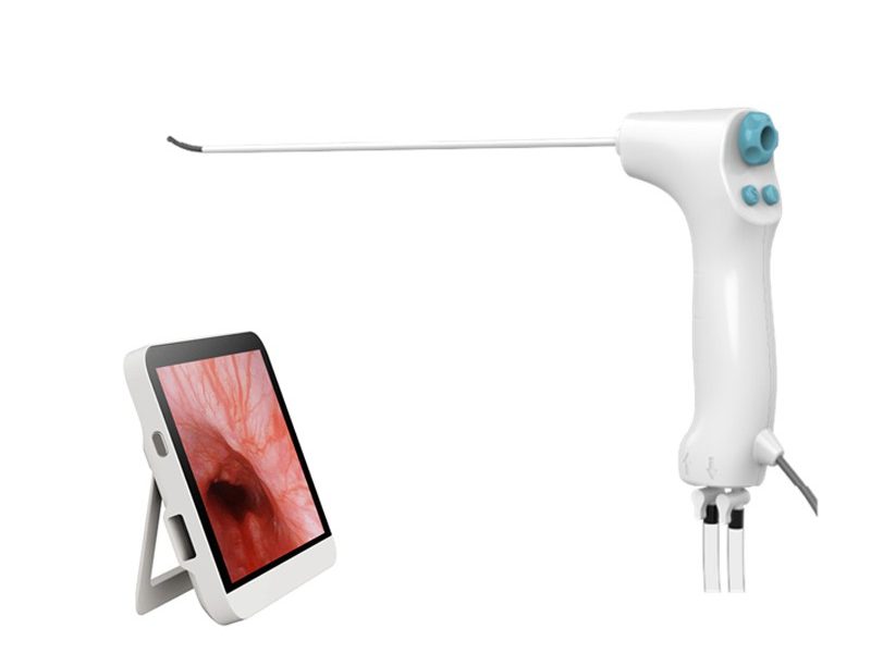
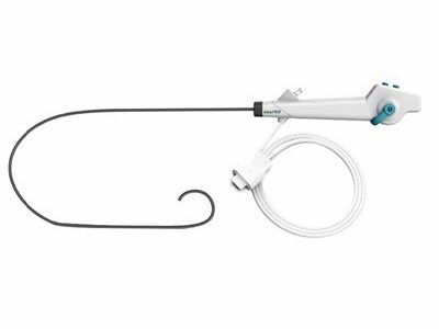
- Scope: The scope is the main body of the hysteroscope. It is a thin, flexible tube that is inserted into the vagina and through the cervix into the uterus. The scope contains a light source, a lens system, and a working channel.
- Light Source: The light source provides illumination inside the uterus, allowing the physician to visualize the uterine cavity clearly. Most portable hysteroscopes use LED lights, which are energy-efficient and provide bright, uniform illumination.
- Lens System: The lens system in the scope magnifies the images of the uterine cavity and transmits them to a display monitor. The quality of the lens system determines the clarity and resolution of the images.
- Working Channel: The working channel is a small tube within the scope that allows the passage of surgical instruments, such as biopsy forceps or scissors. This enables the physician to perform diagnostic and therapeutic procedures during the hysteroscopy.
- Display Monitor: The display monitor is used to view the images captured by the hysteroscope. It provides a real-time visualization of the uterine cavity, allowing the physician to make accurate diagnoses and perform procedures.
Working Principle of a Portable Hysteroscope
The working principle of a portable hysteroscope is based on the concept of direct visualization of the uterine cavity. Here is a step-by-step explanation of how a portable hysteroscope works:
- Insertion: The physician first prepares the patient for the hysteroscopy by performing a pelvic examination and administering a local anesthetic. The scope is then gently inserted through the vagina and into the cervix. The physician carefully guides the scope through the cervical canal and into the uterine cavity.
- Illumination: Once the scope is inside the uterine cavity, the light source is turned on. The LED lights at the tip of the scope illuminate the uterine cavity, making it visible to the physician.
- Image Capture: The lens system in the scope captures the images of the uterine cavity and transmits them to the display monitor. The images are magnified and displayed in real-time, allowing the physician to examine the uterine lining, the fallopian tube openings, and any abnormalities or lesions.
- Diagnostic and Therapeutic Procedures: If the physician identifies any abnormalities during the hysteroscopy, they can use the working channel to insert surgical instruments and perform diagnostic or therapeutic procedures. For example, a biopsy can be taken to obtain a tissue sample for further analysis, or a polyp or fibroid can be removed.
- Removal: After the examination and any necessary procedures are completed, the scope is carefully removed from the uterus and vagina.
Benefits of a Portable Hysteroscope
Portable hysteroscopes offer several advantages over traditional hysteroscopes, including:
- Portability: As the name suggests, portable hysteroscopes are lightweight and easy to transport. They can be used in various settings, such as clinics, offices, or even in the patient’s home. This makes them convenient for both patients and physicians.
- Cost-Effectiveness: Portable hysteroscopes are generally more affordable than traditional hysteroscopes. They do not require a large, dedicated operating room or expensive equipment, which reduces the overall cost of the procedure.
- Patient Comfort: The use of a portable hysteroscope is less invasive and more comfortable for the patient compared to traditional hysteroscopy. The procedure can be performed on an outpatient basis, without the need for general anesthesia, which reduces the risk of complications and allows the patient to recover quickly.
- Real-Time Visualization: Portable hysteroscopes provide real-time visualization of the uterine cavity, allowing the physician to make accurate diagnoses and perform procedures immediately. This improves the efficiency and effectiveness of the treatment.
Types of Portable Hysteroscopes
There are several types of portable hysteroscopes available on the market, each with its own unique features and advantages. Some of the common types include:
- Disposable Hysteroscope: Disposable hysteroscopes are designed for single use only. They eliminate the need for sterilization, which reduces the risk of cross-contamination and saves time and resources.
- Single Use Hysteroscope: Single use hysteroscopes are similar to disposable hysteroscopes, but they may be more durable and can be used for multiple procedures. They are also designed to reduce the risk of cross-contamination.
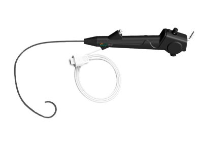
- Flexible Hysteroscope: Flexible hysteroscopes are made of a flexible material that allows them to bend and adapt to the shape of the uterine cavity. This makes them more comfortable for the patient and allows for better visualization of the uterine lining.
Conclusion
In conclusion, a portable hysteroscope is a valuable tool in the field of gynecology. Its working principle is based on direct visualization of the uterine cavity, which allows for accurate diagnoses and effective treatment of various gynecological conditions. The portability, cost-effectiveness, and patient comfort of portable hysteroscopes make them an attractive option for both patients and physicians.


If you are interested in purchasing a portable hysteroscope or have any questions about our products, please feel free to contact us for a consultation. We are committed to providing high-quality medical devices and excellent customer service.
References
- American College of Obstetricians and Gynecologists. (2019). Practice Bulletin No. 194: Hysteroscopy. Obstetrics & Gynecology, 133(6), e214-e230.
- Falcone, T., & Hurd, W. W. (Eds.). (2018). Clinical Gynecologic Endoscopy. Lippincott Williams & Wilkins.
- Magos, A., & Martin-Hirsch, P. L. (2016). Hysteroscopy: A Practical Guide. Springer.

 Blog
Blog
 Blog
Blog
 Blog
Blog