What is the availability of single use cystoscopes in developing countries?
Hey there! As a supplier of Single Use Cystoscopes, I’ve been thinking a lot about the availability of…
Read article →

Uterine imaging plays a crucial role in diagnosing various gynecological conditions, ranging from benign uterine disorders to malignancies. Among the many diagnostic tools available, hysteroscopy and magnetic resonance imaging (MRI) are two commonly used techniques. As a hysteroscope supplier, I have witnessed firsthand the advancements in hysteroscopy technology and understand the nuances of how this method compares to MRI in uterine imaging.
Hysteroscopy is a minimally invasive procedure that allows direct visualization of the uterine cavity. It involves inserting a thin, lighted tube called a hysteroscope through the vagina and cervix into the uterus. This direct access provides real – time images of the uterine lining, allowing healthcare providers to identify and diagnose conditions such as polyps, fibroids, adhesions, and abnormal bleeding.
One of the significant advantages of hysteroscopy is its high level of accuracy in detecting and diagnosing intrauterine pathologies. The ability to directly visualize the uterine cavity enables the identification of even small lesions that may be missed by other imaging modalities. For example, endometrial polyps, which are common causes of abnormal uterine bleeding, can be clearly seen and accurately characterized during hysteroscopy. Additionally, hysteroscopy allows for the simultaneous performance of therapeutic procedures, such as polyp removal or biopsy, which can be done immediately after diagnosis.
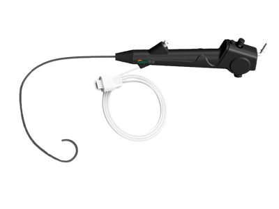
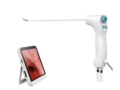
There are different types of hysteroscopes available in the market. Disposable Hysteroscope offers a cost – effective and hygienic solution, eliminating the need for sterilization between uses. This is particularly beneficial in settings where infection control is a major concern. On the other hand, Flexible Hysteroscope provides greater flexibility and maneuverability, allowing for easier navigation through the uterine cavity, especially in cases where the uterine anatomy is complex. Our Flexible Hysteroscope is manufactured with high – quality materials, ensuring clear visualization and reliable performance.
However, hysteroscopy also has its limitations. It is an invasive procedure, which means there is a small risk of complications such as uterine perforation, infection, and bleeding. It also requires specialized training for the healthcare provider to perform the procedure safely and effectively. Moreover, hysteroscopy only provides a view of the uterine cavity and does not provide information about the surrounding pelvic structures.
Magnetic resonance imaging (MRI) is a non – invasive imaging technique that uses a strong magnetic field and radio waves to generate detailed images of the body. In uterine imaging, MRI can provide multi – planar views of the uterus and the surrounding pelvic organs, including the ovaries, fallopian tubes, and pelvic floor muscles.
One of the main advantages of MRI is its ability to provide a comprehensive assessment of the pelvic anatomy. It can accurately depict the size, location, and extent of uterine masses, such as fibroids and adenomyosis. MRI is also useful in evaluating the staging of uterine malignancies, as it can show the involvement of adjacent tissues and lymph nodes. Additionally, MRI does not use ionizing radiation, making it a safer option for patients who may require repeated imaging studies.


MRI is particularly valuable in cases where the diagnosis is unclear or when a more detailed evaluation of the pelvic structures is needed. For example, in patients with suspected deep – infiltrating endometriosis, MRI can help identify the extent of the disease and guide treatment planning.
However, MRI also has some drawbacks. It is a relatively expensive imaging modality and may not be readily available in all healthcare settings. The procedure is time – consuming, usually taking 30 minutes to an hour or more, and requires the patient to lie still inside a narrow tube, which can be uncomfortable for some individuals. MRI is also contraindicated in patients with certain metallic implants, such as pacemakers or cochlear implants, due to the strong magnetic field.
When comparing hysteroscopy and MRI in uterine imaging, several factors need to be considered.
In terms of detecting intrauterine pathologies, hysteroscopy is generally more accurate than MRI. The direct visualization of the uterine cavity allows for the precise identification of lesions, especially small polyps and early – stage endometrial cancers. However, for evaluating the extent of uterine masses and the involvement of surrounding pelvic structures, MRI is superior. It can provide detailed information about the size, location, and relationship of the mass to adjacent tissues, which is crucial for treatment planning.
Hysteroscopy is an invasive procedure, while MRI is non – invasive. The invasiveness of hysteroscopy means that there are associated risks, although these are relatively rare. On the other hand, the non – invasiveness of MRI makes it a more patient – friendly option, especially for those who are reluctant to undergo an invasive procedure.
Hysteroscopy is generally less expensive than MRI and can be performed in an outpatient setting. It is also more widely available, especially in smaller healthcare facilities. MRI, on the other hand, is a costly procedure and may not be accessible in all areas.
One of the unique advantages of hysteroscopy is its therapeutic potential. It allows for the immediate treatment of certain intrauterine conditions, such as polyp removal or biopsy. MRI, on the other hand, is a purely diagnostic tool and does not offer any therapeutic options.
Both hysteroscopy and MRI have their own strengths and limitations in uterine imaging. Hysteroscopy is a valuable tool for direct visualization and treatment of intrauterine pathologies, while MRI provides a comprehensive assessment of the pelvic anatomy. In many cases, these two imaging modalities can complement each other. For example, MRI can be used as a preliminary screening tool to evaluate the overall pelvic anatomy, and hysteroscopy can be performed for a more detailed examination of the uterine cavity and for therapeutic purposes.
As a hysteroscope supplier, I am committed to providing high – quality hysteroscopes that meet the needs of healthcare providers and patients. Our hysteroscopes are designed to offer clear visualization, easy maneuverability, and reliable performance. If you are interested in learning more about our hysteroscope products or would like to discuss procurement options, please feel free to reach out. We look forward to the opportunity to work with you and contribute to better gynecological care.
 Blog
Blog
 Blog
Blog