Are there any cardiac complications associated with bronchoscopy?
Hey there! As a supplier of bronchoscopes, I often get asked about the potential cardiac complications associated with…
Read article →

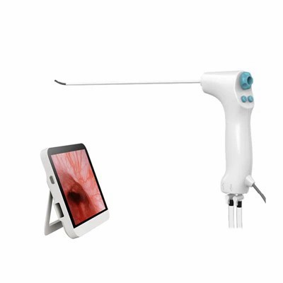
Recurrent pregnancy loss (RPL), defined as two or more consecutive pregnancy losses before the 20th week of gestation, affects approximately 1 – 5% of couples trying to conceive. It is a distressing condition with multiple etiologies, including genetic, anatomical, hormonal, immunological, and thrombophilic factors. Among these, uterine cavity abnormalities are a significant contributing factor, accounting for about 10 – 15% of cases. Evaluating the uterine cavity accurately is crucial for the management of RPL, and a flexible hysteroscope has emerged as a valuable tool in this process.
Uterine cavity abnormalities such as uterine septum, intrauterine adhesions, polyps, and submucosal fibroids can disrupt normal embryo implantation and development, leading to pregnancy loss. Identifying these structural issues is essential because they can often be corrected surgically, potentially improving the chances of a successful pregnancy.
Traditional methods for evaluating the uterine cavity include hysterosalpingography (HSG), transvaginal ultrasound (TVUS), and magnetic resonance imaging (MRI). HSG involves injecting a contrast agent into the uterine cavity and taking X – ray images. While it can provide an overview of the uterine cavity and fallopian tube patency, it has limitations in detecting subtle intrauterine lesions. TVUS is a non – invasive and widely used method, but it may not offer a detailed view of the uterine cavity, especially in cases where the lesions are small or located in the upper part of the uterus. MRI is a more advanced imaging technique, but it is expensive, time – consuming, and not always readily available.
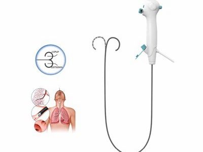
A Flexible Hysteroscope is a thin, flexible endoscope that can be inserted through the cervix into the uterine cavity, allowing direct visualization of the endometrium, uterine walls, and any potential abnormalities. One of the main advantages of a flexible hysteroscope is its flexibility. Unlike rigid hysteroscopes, which may cause discomfort and require more force during insertion, a flexible hysteroscope can easily navigate through the cervical canal and the uterine cavity, adapting to the natural curvature of the uterus. This reduces patient discomfort and the risk of cervical or uterine trauma.
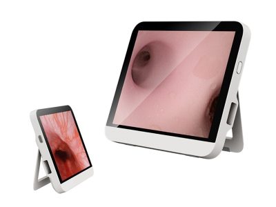
In addition, the high – resolution imaging provided by modern flexible hysteroscopes allows for detailed visualization of the uterine cavity. Small polyps, adhesions, and subtle septa that may be missed by other imaging methods can be clearly identified. The ability to directly visualize the lesions also enables accurate diagnosis and can guide appropriate treatment decisions. For example, if a polyp is detected, it can be biopsied or removed during the same hysteroscopic procedure.
Another benefit of using a flexible hysteroscope is its relatively low – risk nature. The procedure can be performed in an outpatient setting under local anesthesia, which reduces the need for hospitalization and general anesthesia. This not only makes the procedure more convenient for the patient but also lowers the overall cost of the evaluation.
Several studies have demonstrated the effectiveness of flexible hysteroscopy in evaluating the uterine cavity in women with RPL. A study published in the Journal of Minimally Invasive Gynecology found that flexible hysteroscopy detected uterine cavity abnormalities in 32% of women with RPL, compared to 18% detected by HSG. Another research showed that after hysteroscopic correction of uterine cavity abnormalities in women with RPL, the live birth rate increased significantly.
The real – time visualization provided by flexible hysteroscopy also allows for immediate assessment of the treatment outcome. For instance, after the removal of a uterine septum, the surgeon can confirm the completeness of the resection and the integrity of the uterine cavity. This immediate feedback is crucial for ensuring the success of the treatment and improving the chances of a subsequent successful pregnancy.
In addition to flexible hysteroscopes, Disposable Hysteroscope and Portable Hysteroscope are also available in the market. Disposable hysteroscopes offer the advantage of eliminating the need for sterilization between patients, which reduces the risk of cross – contamination. They are also cost – effective for facilities with a low volume of hysteroscopic procedures.
Portable hysteroscopes, on the other hand, are lightweight and easy to transport. They can be used in various settings, including remote areas or in the field. However, they may have some limitations in terms of image quality and functionality compared to larger, more advanced flexible hysteroscopes.
In conclusion, a flexible hysteroscope is a valuable tool for evaluating the uterine cavity in women with recurrent pregnancy loss. Its flexibility, high – resolution imaging, and low – risk nature make it an ideal choice for accurate diagnosis and treatment of uterine cavity abnormalities. By detecting and correcting these abnormalities, the chances of a successful pregnancy can be significantly improved.
If you are a healthcare provider or a medical institution interested in enhancing your diagnostic capabilities for recurrent pregnancy loss, we invite you to explore our range of high – quality Flexible Hysteroscope. Our products are designed with the latest technology to provide the best possible visualization and performance. We also offer Disposable Hysteroscope and Portable Hysteroscope options to meet different clinical needs.


Contact us today to discuss your requirements and start a procurement negotiation. We are committed to providing you with the best products and services to support your clinical practice.
 Blog
Blog
 Blog
Blog