Are there any endoscope monitors with 3D visualization capabilities?
In the dynamic landscape of medical technology, the demand for advanced endoscope monitors with enhanced visualization capabilities has…
Read article →

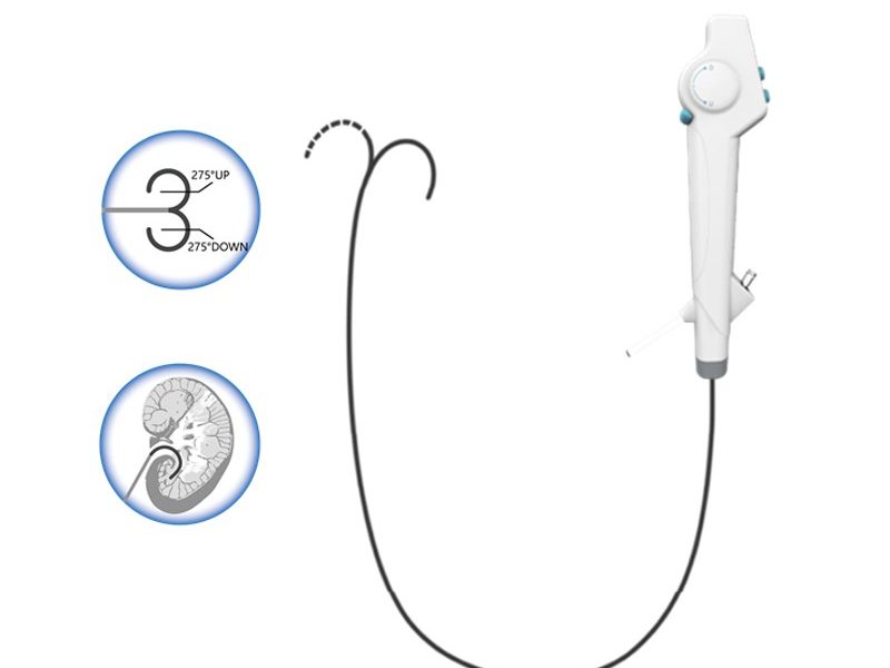
A ureteral tumor can significantly impact a patient’s quality of life and overall health. Ureteroscopy has emerged as a minimally invasive and effective approach for diagnosing and treating ureteral tumors. As a leading ureteroscope supplier, I am excited to share insights into how a ureteral tumor can be ablated using a ureteroscope.
Before the ablation of a ureteral tumor using a ureteroscope, a comprehensive pre – procedure assessment is essential. This includes a detailed medical history review, physical examination, and a series of imaging studies such as CT scans, MRIs, and intravenous pyelograms (IVPs). These tests help in accurately identifying the location, size, and characteristics of the ureteral tumor.
As a ureteroscope supplier, we offer a range of high – quality ureteroscopes, including the Disposable Flexible Ureteroscope. This type of ureteroscope is particularly useful as it can be easily maneuvered through the tortuous ureteral anatomy, reaching the tumor site with precision. Disposable ureteroscopes also eliminate the risk of cross – contamination between patients, which is a crucial consideration in a clinical setting.
The procedure begins with the patient being placed under general or spinal anesthesia. Once the patient is properly anesthetized, a cystoscope is first inserted into the bladder through the urethra. The cystoscope allows the urologist to visualize the bladder and locate the ureteral orifices.
After identifying the ureteral orifice, a guidewire is inserted into the ureter. The guidewire serves as a pathway for the subsequent insertion of the ureteroscope. Our Single Use Ureteroscope is designed to be easily advanced over the guidewire. Its smooth outer surface and flexible design ensure minimal trauma to the ureteral wall during insertion.


The ureteroscope is then carefully advanced along the guidewire into the ureter. As the ureteroscope progresses through the ureter, the urologist can visualize the internal structure of the ureter in real – time. This visualization is crucial for accurately identifying the tumor and planning the ablation strategy.
Once the ureteroscope reaches the tumor site, the urologist can closely examine the tumor. The high – resolution imaging capabilities of our Digital Flexible Ureteroscope provide clear and detailed images of the tumor. This allows the urologist to assess the size, shape, and extent of the tumor, as well as its relationship with the surrounding ureteral tissue.
In addition to visual inspection, the urologist may also perform a biopsy of the tumor. A biopsy forceps can be passed through the working channel of the ureteroscope to obtain a tissue sample for pathological analysis. The pathological diagnosis is essential for determining the appropriate treatment approach, as different types of ureteral tumors may require different ablation techniques.
There are several techniques available for ablating ureteral tumors using a ureteroscope.
Laser ablation is one of the most commonly used techniques. A laser fiber is passed through the working channel of the ureteroscope and positioned at the tumor site. The laser energy is then delivered to the tumor, causing it to vaporize or coagulate. Different types of lasers, such as holmium:YAG lasers, can be used depending on the characteristics of the tumor.
Laser ablation offers several advantages. It is a precise technique that can selectively target the tumor while minimizing damage to the surrounding healthy tissue. It also has a relatively low risk of bleeding, which is important in the delicate ureteral environment.
Electrocoagulation is another option for tumor ablation. An electrocautery probe is passed through the working channel of the ureteroscope. When the probe comes into contact with the tumor, an electrical current is applied, causing the tumor tissue to coagulate and die.
Electrocoagulation is a simple and effective technique, especially for small tumors. However, it may cause more thermal damage to the surrounding tissue compared to laser ablation.
In some cases, mechanical resection may be used. A resectoscope or a basket can be passed through the ureteroscope to physically remove the tumor. This technique is more suitable for larger, pedunculated tumors that can be easily grasped and removed.
After the tumor ablation is completed, the ureteroscope is carefully removed. A ureteral stent may be placed in the ureter to keep it open and facilitate urine drainage. The patient is then transferred to a recovery area for close monitoring.
Post – procedure care includes pain management, antibiotic prophylaxis to prevent infection, and regular follow – up visits. The patient’s urine output, vital signs, and any signs of complications such as bleeding or infection are closely monitored.
Regular follow – up is crucial after ureteral tumor ablation. The patient will typically undergo periodic imaging studies, such as CT scans or IVPs, and cystoscopy to monitor for tumor recurrence. Ureteroscopy may also be repeated to directly visualize the ureteral site.
As a ureteroscope supplier, we understand the importance of providing high – quality instruments for both the initial procedure and follow – up examinations. Our advanced ureteroscopes are designed to meet the needs of urologists in various clinical scenarios, ensuring accurate diagnosis and effective treatment.
Ureteroscopic ablation of ureteral tumors is a minimally invasive and effective treatment option. With the right equipment and a skilled urologist, this procedure can offer patients a better prognosis and a quicker recovery compared to traditional open surgery.
If you are a healthcare provider interested in purchasing high – quality ureteroscopes for your clinical practice, we invite you to contact us for a detailed discussion about our product range and pricing. We are committed to providing the best solutions for ureteral tumor ablation and other urological procedures.
 Blog
Blog
 Blog
Blog