What is the cost of a single use bronchoscope?
Hey there! As a supplier of Single Use Bronchoscopes, I often get asked about the cost of these…
Read article →

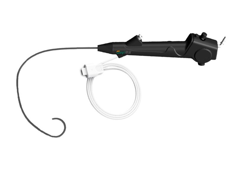
Prostate enlargement, also known as benign prostatic hyperplasia (BPH), is a common condition among aging men. It can lead to various urinary symptoms and complications, often necessitating diagnostic procedures to assess the urinary tract’s condition. One such crucial diagnostic tool is the flexible cystoscope. As a leading supplier of Flexible Cystoscope, I am well – versed in how this device operates and its significance in patients with a history of prostate enlargement.
Before delving into how a flexible cystoscope works, it’s essential to understand the implications of prostate enlargement. The prostate gland, which is located just below the bladder and surrounds the urethra, can gradually increase in size as men age. This growth can compress the urethra, leading to urinary problems such as difficulty starting urination, weak urine stream, frequent urination, and the feeling of incomplete bladder emptying.
In more severe cases, prostate enlargement can cause urinary retention, bladder stones, and even kidney damage. To accurately diagnose these issues and plan appropriate treatment, doctors need to visualize the inside of the urinary tract, including the bladder and the urethra. This is where the flexible cystoscope comes into play.
A flexible cystoscope is a thin, tube – like instrument equipped with a light source and a camera at its tip. It is designed to be inserted into the urethra and advanced into the bladder. The flexibility of the device allows it to navigate the natural curves of the urinary tract, minimizing discomfort to the patient.
The cystoscope consists of several key components. The outer sheath protects the internal components and provides a smooth surface for insertion. Inside the sheath, there are channels for various functions. One channel is used for the light guide, which delivers light from an external source to the tip of the cystoscope, illuminating the inside of the urinary tract. Another channel houses the fiber – optic bundle or digital camera that captures images and transmits them to an external monitor.
There are also channels for irrigation and instrument insertion. Irrigation is necessary to keep the field of view clear by flushing out blood, mucus, or debris. The instrument channel allows the doctor to pass additional tools, such as biopsy forceps or laser fibers, for diagnostic or therapeutic purposes.
Before the cystoscopy procedure, the patient is typically given local anesthesia in the form of a gel applied to the urethra. This helps to numb the area and reduce discomfort during insertion. The patient is then positioned on an examination table, usually lying on their back with their knees bent and feet apart.
The doctor carefully inserts the flexible cystoscope into the urethral opening at the tip of the penis (in men). As the cystoscope is advanced through the urethra, the doctor can use the control knobs on the handle to bend the tip of the cystoscope, allowing it to follow the natural curvature of the urethra. In patients with prostate enlargement, the urethra may be narrowed or distorted due to the enlarged prostate gland. The flexibility of the cystoscope enables it to navigate these changes more easily compared to a rigid cystoscope.
Once the cystoscope reaches the bladder, the doctor can examine the bladder lining, the openings of the ureters (the tubes that carry urine from the kidneys to the bladder), and the area where the urethra exits the bladder. In patients with prostate enlargement, the doctor can assess the degree of obstruction caused by the enlarged prostate. They can look for signs of bladder wall changes, such as trabeculation (thickening and ridges in the bladder wall), which may indicate long – standing urinary obstruction.
If any suspicious areas are identified during the examination, the doctor can use the instrument channel to pass biopsy forceps and obtain tissue samples for further analysis. This is important for ruling out the presence of bladder cancer, which can sometimes co – exist with prostate enlargement.
In addition to diagnosis, the flexible cystoscope can also be used for some therapeutic procedures. For example, if there are small bladder stones, the doctor can use a laser fiber passed through the instrument channel to break up the stones into smaller pieces, which can then be flushed out of the bladder.
After the examination and any necessary procedures are completed, the cystoscope is carefully removed from the urethra. The patient may experience some mild discomfort or a burning sensation during urination for a short period after the procedure, but this usually resolves within a day or two.
Compared to more invasive surgical procedures, cystoscopy using a flexible cystoscope is a minimally invasive option. It can be performed in an outpatient setting, and patients can usually return home the same day. This reduces the risk of complications associated with general anesthesia and major surgery.
The high – quality images provided by the flexible cystoscope allow doctors to accurately assess the condition of the urinary tract. They can detect early signs of problems such as bladder cancer, inflammation, or strictures, which may not be apparent on other imaging tests.
The flexibility of the cystoscope reduces the discomfort experienced by the patient during the procedure. It can adapt to the natural shape of the urinary tract, minimizing the need for forceful manipulation.


In addition to the flexible cystoscope, we also offer Portable Cystoscope and Single Use Cystoscope. Portable cystoscopes are lightweight and easy to transport, making them suitable for use in different clinical settings, such as in remote areas or during bedside examinations. Single – use cystoscopes eliminate the need for reprocessing, reducing the risk of cross – contamination and ensuring a high level of patient safety.
The flexible cystoscope is an invaluable tool in the diagnosis and management of patients with a history of prostate enlargement. Its ability to navigate the complex anatomy of the urinary tract, provide clear visualization, and support both diagnostic and therapeutic procedures makes it an essential part of urological practice.
As a supplier of high – quality flexible cystoscopes, we are committed to providing healthcare professionals with reliable and innovative devices. If you are interested in learning more about our products or are looking to purchase cystoscopes for your medical facility, we invite you to reach out for a procurement discussion. We can provide detailed product information, pricing, and support to meet your specific needs.
 Blog
Blog
 Blog
Blog
 Blog
Blog