What are the latest advancements in ureteroscope technology?
In the realm of urological surgery, ureteroscopes have long been indispensable tools, enabling minimally invasive procedures for the…
Read article →

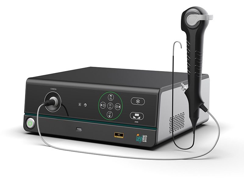
How does an endoscope camera system work?
As a seasoned supplier of endoscope camera systems, I’ve witnessed firsthand the transformative power of this technology in the medical field. Endoscope camera systems are essential tools that allow medical professionals to visualize the internal organs and structures of the human body without the need for large – scale invasive surgeries. In this blog, I’ll delve into the inner workings of an endoscope camera system, exploring its components, functions, and the processes that make it such a valuable asset in modern medicine.
An endoscope camera system is composed of several key components, each playing a crucial role in its operation.
The endoscope is the most visible part of the system. It is a long, thin tube that is inserted into the body through natural openings such as the mouth, anus, or through small incisions in laparoscopic procedures. There are different types of endoscopes, including rigid and flexible ones. Rigid endoscopes are typically used for procedures in areas where a straight – line view is sufficient, such as the bladder or joints. Flexible endoscopes, on the other hand, can bend and navigate through the winding pathways of the digestive tract, respiratory system, and other complex internal structures.
The endoscope contains a bundle of optical fibers or a digital imaging sensor at its tip. Optical fibers are used in traditional endoscopes to transmit light from a light source at the proximal end of the endoscope to the tip, illuminating the area of interest inside the body. Digital endoscopes, which are becoming more prevalent, use a small camera chip (such as a charge – coupled device or complementary metal – oxide – semiconductor sensor) at the tip to capture images.
A high – quality light source is essential for clear visualization inside the body. The light source provides bright, white light that is transmitted through the endoscope to the area being examined. Xenon lamps were commonly used in the past due to their high – intensity and color – rendering properties. However, more recently, light – emitting diodes (LEDs) have gained popularity. LEDs are more energy – efficient, have a longer lifespan, and can provide a more consistent light output. The light is directed through the optical fibers or other light – guiding mechanisms in the endoscope to illuminate the target area.
The camera at the tip of the endoscope captures the images of the internal structures. These images are then transmitted to the image processing unit. In digital endoscopes, the image data is sent electronically via cables or wirelessly. The image processing unit enhances the captured images, adjusting parameters such as brightness, contrast, and color balance. It also can perform functions like image stitching in some advanced systems, which combines multiple images to create a larger, more comprehensive view.
The monitor is where the processed images are displayed. Medical professionals can view the internal structures in real – time on the monitor, allowing them to make accurate diagnoses and perform procedures. High – resolution monitors are preferred to ensure that fine details of the tissues and organs are visible. Some monitors also support features like picture – in – picture, which allows for simultaneous viewing of different areas or different types of imaging (e.g., normal and enhanced imaging modes).
When a medical procedure begins, the endoscope is carefully inserted into the body. The medical professional uses the controls on the endoscope’s handle to navigate it through the internal passages. In the case of a flexible endoscope, the controls can be used to bend the tip of the endoscope in different directions, allowing it to reach areas that are difficult to access. For example, when performing an upper gastrointestinal endoscopy, the endoscope is inserted through the mouth and guided down the esophagus, stomach, and into the duodenum.
Once the endoscope is in the desired position, the camera at the tip starts capturing images. In a digital endoscope, the camera chip converts the light reflected from the internal tissues into electrical signals. These signals are then processed into digital image data. In an optical fiber – based endoscope, the light reflected from the tissues is transmitted back through the optical fibers to an eyepiece or a camera adapter, where it is then converted into an image.
The captured image data is sent to the image processing unit. The unit analyzes the data and applies various algorithms to enhance the image quality. For instance, it can reduce noise, improve sharpness, and adjust the color to make the tissues appear more natural. This processed image is then sent to the monitor for display.
The medical professional views the images on the monitor and can identify any abnormalities such as tumors, ulcers, or inflammation. Based on the findings, they can decide on the appropriate course of action, which may include taking biopsies, performing minor surgical procedures, or prescribing medications. For example, during a colonoscopy, if a polyp is detected, the medical professional can use specialized tools passed through the working channel of the endoscope to remove it.
Modern endoscope camera systems come with a range of advanced features that enhance their functionality and diagnostic capabilities.
The demand for high – definition and 4K imaging in endoscopy is increasing. High – resolution imaging provides more detailed views of the tissues, allowing for earlier detection of diseases. Our 4K Medical Endoscope Camera System offers exceptional image quality, with sharp details and accurate color representation. This is particularly important in detecting early – stage cancers and other subtle abnormalities.
Narrow – Band Imaging is a technique that uses specific wavelengths of light to enhance the visualization of the blood vessels and surface patterns of the tissues. By highlighting these features, NBI can help in differentiating between normal and abnormal tissues more easily. For example, it can make precancerous lesions more visible, which may not be as apparent under normal white – light imaging.
Some endoscope camera systems support fluorescence imaging. In this technique, fluorescent dyes are injected into the patient’s body, and the endoscope is equipped with a light source that can excite the dyes. The fluorescent light emitted by the dyes can reveal specific molecular or cellular characteristics of the tissues. This can be useful in detecting cancer cells or in mapping the boundaries of tumors more accurately.
There are different types of endoscope camera systems tailored to specific medical applications.
These systems are designed for a wide range of general endoscopy procedures, such as upper gastrointestinal endoscopy, colonoscopy, and bronchoscopy. They are versatile and can be used in different clinical settings, from outpatient clinics to large hospitals.
Laparoscopic camera systems are used in minimally invasive surgical procedures. They are typically rigid endoscopes and are inserted through small incisions in the abdomen. These systems provide a clear view of the abdominal organs during procedures like laparoscopic cholecystectomy (removal of the gallbladder) or laparoscopic appendectomy.
As a supplier of endoscope camera systems, we offer high – quality products that are reliable and innovative. Our systems are designed with the latest technology, providing excellent image quality and advanced features to meet the needs of medical professionals. We have a team of experts who can provide technical support and training to ensure that our customers can make the most of our products.


If you are in the market for an endoscope camera system, whether it’s for a small clinic or a large – scale hospital, we invite you to contact us for a detailed discussion. Our products are designed to enhance the accuracy of diagnoses and the effectiveness of medical procedures. Let’s work together to improve patient care with our state – of – the – art endoscope camera systems. Reach out to us to start the procurement process and explore how our solutions can fit your specific requirements.
 Blog
Blog
 Blog
Blog
 Blog
Blog