Top 10 ENT Suppliers in China
Introduction to ENT ENT, which stands for Ear, Nose, and Throat, is a specialized medical field dealing with…
Read article →

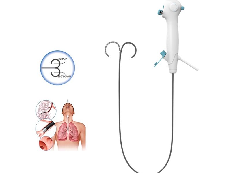
As a supplier of Single Use Bronchoscopes, I am often asked about the main components of these essential medical devices. In this blog post, I will delve into the key parts that make up a single use bronchoscope, highlighting their functions and importance in the field of respiratory medicine.
The insertion tube is the long, flexible part of the bronchoscope that is inserted into the patient’s airway. It is typically made of a durable, biocompatible material such as polyurethane or silicone, which ensures patient safety and comfort during the procedure. The insertion tube is designed to be thin and flexible, allowing it to navigate through the complex anatomy of the respiratory tract with minimal trauma to the patient.
The outer diameter of the insertion tube can vary depending on the specific model and intended use of the bronchoscope. In general, smaller diameter tubes are used for pediatric patients or for accessing smaller airways, while larger diameter tubes may be used for adult patients or for procedures that require the passage of additional instruments.
The insertion tube also contains several channels that serve different functions. One of the most important channels is the working channel, which allows for the passage of various instruments such as biopsy forceps, brushes, and suction catheters. This enables the physician to perform diagnostic and therapeutic procedures, such as taking tissue samples for biopsy or removing foreign bodies from the airway.
Another channel in the insertion tube is the light guide channel, which transmits light from the light source at the proximal end of the bronchoscope to the distal tip. This illuminates the airway, allowing the physician to visualize the internal structures clearly. Additionally, there may be a channel for the delivery of water or air, which can be used to clean the lens or to inflate the airway for better visualization.
The control section is the part of the bronchoscope that is held by the physician during the procedure. It contains a series of controls and knobs that allow the physician to manipulate the insertion tube and perform various functions.
One of the main controls on the control section is the deflection control, which allows the physician to bend the distal tip of the insertion tube in different directions. This is essential for navigating through the complex anatomy of the airway and for accessing different areas of the lungs. The deflection control typically consists of two knobs or levers, which can be used to bend the tip up and down or left and right.
The control section also contains controls for the working channel, such as the suction control and the instrument insertion port. The suction control allows the physician to apply suction through the working channel, which can be used to remove secretions or debris from the airway. The instrument insertion port is where the physician inserts the various instruments into the working channel.
In addition to these controls, the control section may also contain buttons or switches for other functions, such as the light intensity control, the camera focus control, and the image capture button. These controls allow the physician to adjust the settings of the bronchoscope and to capture images or videos of the airway for documentation and diagnostic purposes.
The light source is an essential component of the bronchoscope, as it provides the illumination necessary for visualizing the airway. Most single use bronchoscopes are equipped with a built-in light source, which is typically a light-emitting diode (LED). LEDs are preferred over traditional light sources, such as halogen bulbs, because they are more energy-efficient, produce less heat, and have a longer lifespan.
The light source is connected to the bronchoscope through a light guide cable, which transmits the light from the source to the distal tip of the insertion tube. The light guide cable is made of a bundle of optical fibers, which are capable of transmitting light over long distances with minimal loss of intensity.
The intensity of the light can be adjusted using the light intensity control on the control section of the bronchoscope. This allows the physician to optimize the illumination based on the specific conditions of the procedure and the patient’s airway.
The camera and imaging system are responsible for capturing images and videos of the airway during the bronchoscopy procedure. Most single use bronchoscopes are equipped with a high-resolution video camera at the distal tip of the insertion tube. The camera is typically a charge-coupled device (CCD) or a complementary metal-oxide-semiconductor (CMOS) sensor, which can capture clear and detailed images of the internal structures of the airway.
The images and videos captured by the camera are transmitted through a cable to the imaging system, which is usually a monitor or a computer. The imaging system allows the physician to view the real-time images of the airway on a screen, which can be used for diagnostic and therapeutic purposes. The imaging system may also have features such as image enhancement, measurement tools, and the ability to store and print images and videos for documentation.
Some single use bronchoscopes may also be equipped with additional imaging modalities, such as narrow band imaging (NBI) or autofluorescence imaging (AFI). These techniques can provide additional information about the tissue characteristics, which can be useful for detecting early-stage cancers or other abnormalities.
The proximal connector is the part of the bronchoscope that connects to the external devices, such as the light source, the video processor, and the suction unit. It typically consists of a series of connectors and ports, which are designed to be compatible with the specific devices used in the bronchoscopy procedure.
The proximal connector is an important part of the bronchoscope, as it ensures the proper functioning of the device and the transmission of signals between the different components. It is important to ensure that the proximal connector is properly connected and sealed to prevent any leakage of fluids or air.
In conclusion, a single use bronchoscope is a complex medical device that consists of several main components, each of which plays a crucial role in its function. The insertion tube, control section, light source, camera and imaging system, and proximal connector all work together to enable the physician to visualize the airway, perform diagnostic and therapeutic procedures, and obtain valuable information about the patient’s respiratory health.
As a supplier of Single Use Bronchoscopes, we are committed to providing high-quality products that meet the needs of our customers. Our single use bronchoscopes are designed to be safe, reliable, and easy to use, and they are equipped with the latest technology and features to ensure optimal performance.


If you are interested in learning more about our Single Use Bronchoscopes or if you have any questions or inquiries, please do not hesitate to contact us. We would be happy to discuss your specific requirements and to provide you with more information about our products and services. We look forward to the opportunity to work with you and to help you meet your medical needs.
 Blog
Blog
 Blog
Blog
 Blog
Blog