What are the types of bronchoscopes?
When it comes to diagnosing and treating various respiratory conditions, bronchoscopes play a pivotal role. As a professional…
Read article →

Hey there! As a cystoscope supplier, I’ve seen firsthand how these nifty devices play a crucial role in detecting urethral tumors. So, I thought I’d share some insights on what signs doctors look for when using a cystoscope to spot these tumors.


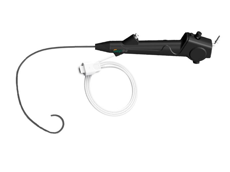
First off, let’s quickly talk about what a cystoscope is. It’s a thin, tube – like instrument with a light and a camera at the end. Doctors insert it through the urethra to get a direct view of the inside of the urethra and the bladder. There are different types of cystoscopes, like the Disposable Flexible Cystoscope, Flexible Cystoscope, and Disposable Cystoscope. Each has its own advantages, but they all serve the same main purpose: to help diagnose problems in the urinary tract.
One of the most obvious signs that doctors look for is the presence of abnormal growths. Urethral tumors can have different visual characteristics.
When using a cystoscope, a doctor might see small or large masses protruding from the urethral wall. These masses can be smooth – surfaced or have an irregular, bumpy appearance. Small nodules might be just a few millimeters in size, while larger masses can be several centimeters across. For example, a papillary tumor often looks like a cauliflower – shaped growth sticking out into the urethral lumen. It’s kind of like seeing a little alien growth in there!
The color of the tumor can also give important clues. Normal urethral tissue is usually a pale pink color. Tumors, on the other hand, can be a variety of colors. They might be white, which could indicate areas of dead or abnormal cells. Sometimes, they can be red or purple, suggesting increased blood flow to the area, which is common in growing tumors. Dark – colored tumors might be a sign of bleeding or the presence of certain types of cancer cells.
Another tell – tale sign is the presence of ulcers. An ulcer is an open sore on the urethral wall. Tumors can cause the tissue to break down, forming these ulcers. Ulcers often have a ragged edge and a base that might look different from the surrounding healthy tissue. They can be painful for the patient, and the doctor can see the raw, exposed area when looking through the cystoscope.
The cystoscope also allows doctors to closely examine the texture and structure of the urethral wall.
Urethral tumors can cause the wall of the urethra to thicken. Instead of the normal thin and smooth appearance, the wall might look and feel thicker. This thickening can be focal, meaning it’s only in one area, or it can be more widespread. Doctors use the cystoscope to measure the thickness of the wall in different parts of the urethra and compare it to what’s considered normal.
The normal urethral wall is relatively smooth. But when there’s a tumor, the wall can become irregular. There might be bumps, ridges, or indentations that are not supposed to be there. These irregularities can be a sign that the tumor is invading the surrounding tissue and changing its structure.
The lumen is the hollow space inside the urethra through which urine flows. Tumors can have a significant impact on this space.
As a tumor grows, it can narrow the urethral lumen. This is called stricture. The doctor can see that the normal open space is getting smaller, and it might be more difficult for the cystoscope to pass through. Narrowing of the lumen can also cause problems with urine flow, leading to symptoms like difficulty urinating, weak urine stream, or a feeling of incomplete emptying of the bladder.
In severe cases, a large tumor can completely block the urethral lumen. When this happens, urine can’t flow out properly, and the patient might experience severe pain and swelling in the bladder. The cystoscope can clearly show the point where the blockage is occurring and give the doctor an idea of how to treat it.
Urethral tumors often cause inflammation in the surrounding tissue.
The doctor can see signs of inflammation, such as redness, swelling, and an increased number of blood vessels in the area. Inflammatory cells are the body’s way of trying to fight off the abnormal growth. The cystoscope can pick up these subtle changes in the tissue, which can be an early sign of a tumor.
Bleeding is another common sign. Tumors can be fragile, and even minor trauma during the cystoscopy procedure or normal urine flow can cause them to bleed. The doctor might see small streaks of blood in the urine or actual bleeding from the tumor site. This bleeding can be a sign of a more aggressive tumor or one that has invaded blood vessels.
As a cystoscope supplier, I know that using the right type of cystoscope is crucial for accurate detection of urethral tumors. The Disposable Flexible Cystoscope is great because it’s single – use, which reduces the risk of cross – contamination. It’s also flexible, so it can easily navigate the curves of the urethra without causing too much discomfort to the patient.
The Flexible Cystoscope offers more flexibility and can be reused after proper sterilization. It has high – quality optics, which means the doctor can get a clear and detailed view of the urethral and bladder interior.
The Disposable Cystoscope is a cost – effective option for clinics and hospitals. It’s pre – sterilized and ready to use, saving time and effort in the sterilization process.
If you’re in the medical field and are looking for reliable cystoscopes for detecting urethral tumors or other urinary tract problems, we’ve got you covered. Our cystoscopes are designed with the latest technology to provide accurate and clear images, helping doctors make better diagnoses.
Whether you’re a urologist in a large hospital or a doctor in a small clinic, having the right cystoscope can make a big difference in your practice. If you’re interested in learning more about our cystoscopes or want to discuss a potential purchase, don’t hesitate to reach out. We’re always happy to talk about how our products can meet your needs.
 Blog
Blog
 Blog
Blog