Can a hysteroscopy be used to remove intrauterine foreign bodies?
Can a hysteroscopy be used to remove intrauterine foreign bodies? Intrauterine foreign bodies can pose significant health risks…
Read article →

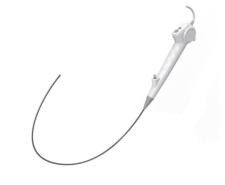
As a supplier of choledochoscopes, I’ve had the chance to see firsthand how these nifty devices help doctors peer into the biliary and pancreatic ducts. They’re super useful, but like any medical tool, they’ve got their limitations. Let’s dig into what those are when it comes to using a choledochoscope to visualize the pancreatic duct.
First off, the anatomy of the pancreatic duct is a real challenge. The pancreatic duct is often smaller and more tortuous compared to the bile duct. Choledochoscopes are generally designed with the biliary system in mind, and their size and flexibility might not be the best fit for the pancreatic duct. The diameter of the scope can be too large to easily navigate through the narrow pancreatic duct. This means that in some cases, it can be tough to get the scope all the way to the areas of interest. For example, if there’s a small branch of the pancreatic duct that’s suspected of having a problem, the choledochoscope might not be able to reach it due to its size. You can check out our Single Use Choledochoscope to see the specifications and how it might work in different anatomical situations.


Another limitation is the image quality. While choledochoscopes have come a long way in terms of providing clear visuals, when it comes to the pancreatic duct, the image can still be less than ideal. The pancreatic duct has a different structure and composition compared to the bile duct. The mucus and secretions in the pancreatic duct can cloud the view, making it hard to get a crisp image. Also, the angle at which the scope can view the pancreatic duct might not be optimal. Sometimes, the duct might be oriented in such a way that the scope can only get a partial or distorted view of the area. This can make it difficult for doctors to accurately diagnose conditions like tumors or strictures. Our Single Use Choledochoscope is designed to improve image quality, but there are still these inherent challenges with the pancreatic duct.
Manipulation is also a big issue. The pancreatic duct is located deep within the body, and getting the choledochoscope to the right position and then maneuvering it to get a good look around can be a real pain. The scope needs to be steered through a complex network of ducts and organs. There’s a risk of causing trauma to the pancreatic duct or surrounding tissues during the manipulation process. If the scope is forced too hard or in the wrong direction, it can lead to complications like pancreatitis or bleeding. Doctors need to have a high level of skill and experience to use the choledochoscope safely and effectively in the pancreatic duct. Our Disposable Choledochoscope is designed to be more user – friendly, but the manipulation challenges still remain.
In addition, the diagnostic capabilities are somewhat limited. Just being able to see the inside of the pancreatic duct isn’t always enough to make a definite diagnosis. For example, some early – stage tumors or precancerous lesions might not be easily distinguishable from normal tissue. The choledochoscope can only provide a visual inspection, and it can’t perform in – depth tissue analysis on the spot. To confirm a diagnosis, additional tests like biopsies or laboratory analyses are usually required. This means that the choledochoscope is just one part of the diagnostic process and might not give a complete picture on its own.
There are also some issues related to the environment inside the pancreatic duct. The pancreatic juice has a unique chemical composition, and it can have an impact on the performance of the choledochoscope. Over time, the juice can corrode or damage the scope’s components, especially if the scope isn’t properly designed to withstand these conditions. This can lead to a shorter lifespan of the scope and more frequent replacements.
Now, despite these limitations, choledochoscopes still play an important role in visualizing the pancreatic duct. They can provide valuable information that might not be available through other imaging techniques. And at our company, we’re constantly working on improving our products to overcome these challenges. We’re researching new materials to make the scopes more flexible and resistant to the pancreatic environment. We’re also looking into ways to enhance the image quality and diagnostic capabilities.
If you’re in the medical field and are interested in using our choledochoscopes for pancreatic duct visualization, we’d love to have a chat with you. Whether you’re a hospital, a clinic, or a medical research institution, we can discuss how our products can fit your needs. Contact us to start the procurement process and let’s see how we can work together to improve patient care.
References
 Blog
Blog
 Blog
Blog