Blog

Can an Endoscope System be used for endoscopic variceal ligation?

Endoscopic variceal ligation (EVL) is a well – established therapeutic procedure for the management of esophageal varices, which are enlarged veins in the esophagus often associated with liver cirrhosis and portal hypertension. As a leading supplier of endoscope systems, I am frequently asked whether our endoscope systems can be used for endoscopic variceal ligation. In this blog, I will delve into the details of this question, exploring the functionality, suitability, and benefits of our endoscope systems in the context of EVL.

Understanding Endoscopic Variceal Ligation

Before discussing the use of endoscope systems for EVL, it is essential to understand what the procedure entails. Esophageal varices occur when the normal blood flow through the liver is obstructed, usually due to liver disease. As a result, blood is redirected through smaller veins in the esophagus, causing them to enlarge and become vulnerable to rupture, which can lead to life – threatening bleeding.

EVL is a minimally invasive procedure that involves placing small rubber bands around the varices using an endoscope. These bands cut off the blood supply to the varices, causing them to shrink and eventually fall off. This reduces the risk of bleeding and can prevent future hemorrhages.

The Role of Endoscope Systems in EVL

An endoscope is a flexible tube with a light and a camera at the end, which allows doctors to visualize the inside of the esophagus, stomach, and other parts of the digestive tract. For EVL, the endoscope serves as the primary tool for both diagnosis and treatment.

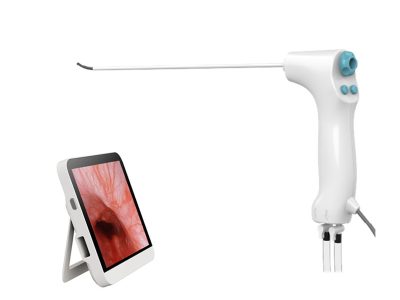
Our endoscope systems are designed to provide high – quality visualization, which is crucial for accurately identifying and targeting the varices. The Video Endoscopy System offers clear and detailed images, enabling doctors to precisely place the rubber bands around the varices. The high – resolution video capabilities ensure that even small varices can be detected and treated effectively.

Key Features of Our Endoscope Systems for EVL

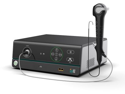
  1. High – Resolution Imaging: Our Endoscope Image Processor is equipped with advanced technology that enhances the clarity and sharpness of the images. This is essential for distinguishing between normal tissue and varices, as well as for guiding the ligation procedure. The processor can adjust the color, contrast, and brightness of the images in real – time, providing optimal visualization under different lighting conditions.
  2. Flexibility and Maneuverability: The endoscopes in our systems are highly flexible, allowing them to navigate the complex anatomy of the esophagus and reach the varices easily. This flexibility reduces the discomfort for the patient and increases the success rate of the procedure. The endoscopes can be bent and rotated in multiple directions, enabling doctors to access varices in difficult – to – reach locations.
  3. Compatibility with Ligation Devices: Our endoscope systems are designed to be compatible with a wide range of ligation devices. This means that doctors can choose the most suitable device for each patient, depending on the size and location of the varices. The endoscopes have a working channel through which the ligation device can be inserted, allowing for seamless integration of the diagnostic and therapeutic aspects of the procedure.
  4. Ergonomic Design: The control handles of our endoscopes are ergonomically designed to fit comfortably in the doctor’s hand. This reduces fatigue during long procedures and allows for precise control of the endoscope. The buttons and switches on the control handle are easily accessible, enabling doctors to operate the endoscope and the ligation device simultaneously.

Benefits of Using Our Endoscope Systems for EVL

  1. Improved Patient Outcomes: By providing clear visualization and precise targeting, our endoscope systems can improve the success rate of EVL. This means that more varices can be effectively treated, reducing the risk of bleeding and improving the patient’s overall prognosis. In addition, the minimally invasive nature of the procedure, facilitated by our high – quality endoscopes, results in less pain and a shorter recovery time for the patient.
  2. Cost – Effectiveness: Our endoscope systems are designed to be durable and reliable, reducing the need for frequent replacements. This can result in cost savings for healthcare providers in the long run. Moreover, the high success rate of EVL using our systems can reduce the need for more invasive and expensive treatments, such as surgery.
  3. Enhanced Safety: The advanced features of our endoscope systems, such as high – resolution imaging and precise maneuverability, contribute to the safety of the EVL procedure. Doctors can better assess the risks and complications associated with the procedure and take appropriate measures to prevent them. The compatibility with ligation devices also ensures that the procedure is performed accurately, minimizing the risk of injury to the patient.

Case Studies

To illustrate the effectiveness of our endoscope systems in EVL, let’s look at a few case studies.

Case 1: A 55 – year – old male with liver cirrhosis and esophageal varices was referred for EVL. Using our Endoscopy Camera System, the doctor was able to clearly visualize the varices in the lower esophagus. The high – resolution images allowed for precise placement of the rubber bands, and the procedure was completed successfully without any complications. The patient’s varices shrank significantly after the procedure, and there was no evidence of bleeding during the follow – up period.

Endoscope Image ProcessorVideo Endoscopy System

Case 2: A 62 – year – old female with a history of variceal bleeding was scheduled for EVL. Our endoscope system, with its flexible and maneuverable endoscope, was able to reach the varices in the upper esophagus, which were difficult to access. The doctor was able to place the rubber bands accurately, and the patient experienced no further bleeding. The patient’s quality of life improved significantly after the procedure, and she was able to resume her normal activities.

Conclusion

In conclusion, our endoscope systems are well – suited for endoscopic variceal ligation. The high – resolution imaging, flexibility, compatibility with ligation devices, and ergonomic design make them an ideal choice for healthcare providers performing this important procedure. By using our endoscope systems, doctors can improve patient outcomes, reduce costs, and enhance the safety of the EVL procedure.

If you are interested in learning more about our endoscope systems for EVL or would like to discuss a potential purchase, please feel free to contact us. We are committed to providing the highest quality medical equipment and excellent customer service.
References

  1. Garcia – Tsao G, Sanyal AJ, Grace ND, Carey WD. Prevention and management of gastroesophageal varices and variceal hemorrhage in cirrhosis. Hepatology. 2007;46(3):922 – 938.
  2. Sarin SK, Lahoti D, Saxena SP, Murthy NS, Makwana UK. Prevalence, prediction, and prognosis of esophageal varices in cirrhosis: a long – term follow – up study. Hepatology. 1992;16(4):1042 – 1048.
  3. Bosch J, Berzigotti A, Piqueras M, et al. Endoscopic variceal ligation versus endoscopic sclerotherapy: a meta – analysis. Hepatology. 1995;22(1):167 – 173.

Published by Jumping Spider™ — single-use endoscopy brand of Zhejiang Geyi Medical Devices Co., Ltd.

Need distributor or OEM information?

Tell us your specialty and target markets — our team will respond with the right documentation and product match.

Send inquiry